
Clinical History
A 65-year-old man presented with confusion, memory loss, and ataxia. His family noted that the onset of memory loss had become noticeable over the past three months and had become particularly striking in the past week when he was noted to exhibit abnormal “sudden, jerking movements.” There was no history of alcoholism, psychiatric disease, lithium exposure, or heavy metal exposure. A CT scan was normal. A biopsy was obtained.
Tissue Site
Brain
Whole Slide Image
The whole slide image provided is an H&E stain from a brain biopsy.
Questions
- The presence of a perivascular inflammatory infiltrate in the biopsy would rule out which of the following?
- Acute demyelinating encephalomyelitis
- Creutzfeldt-Jakob disease
- Lyme disease
- Multiple sclerosis
- Systemic lupus erythematosus
- The presence of vacuolation of the white matter would implicate which of the following?
- Cerebral amyloid angiopathy
- Chronic traumatic encephalopathy
- Parkinson disease
- Progressive supranuclear palsy
- Processing artifact
- In contrast to other dementing diseases, which anatomic site is typically spared in Creutzfeldt-Jakob disease?
- Caudate nucleus
- Cerebellum
- Frontal Lobe
- Hippocampus
- Thalamus
Discussion and Diagnosis
The diagnosis is Creutzfeldt-Jakob disease (CJD), a spongiform encephalopathy…
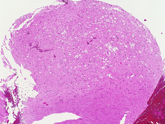
Image A: H&E stain, low magnification. |
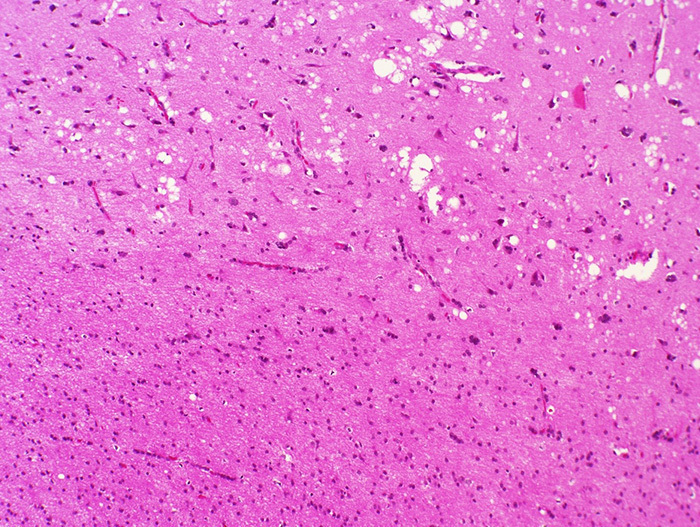
Image B: H&E stain, intermediate magnification. |
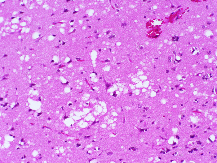
Image C: H&E stain, high magnification. |
Prion protein (PrPc) is a normal protein (the superscripted ‘c’ refers to ‘cellular’) derived from the human prion protein gene (PRNP)…
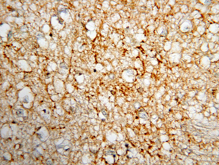
Image D: IHC. Human prion protein or PrP (3F4) after proteinase K pretreatment. |
Electroencephalographic (EEG) studies that exhibit characteristic periodic triphasic complexes at about one per second can be found in the majority of cases, and MRI findings of T2-hyperintensity in basal ganglia and thalami with persistent restricted diffusion on diffusion-weighted imaging (DWI) have been incorporated into the diagnostic criteria for sporadic CJD. Combined with an appropriate clinical examination and cerebrospinal fluid studies demonstrating elevated 14-3-3 protein level, the diagnosis is typically made without a biopsy. However, in certain cases, the disease can be suspected in the absence of typical findings. In such cases, special handling of the brain sample is strongly urged to avoid contamination of laboratory instruments, surfaces, and personnel. The CDC has published guidelines on the proper handling of these samples and familiarity with the guidelines is essential prior to handling a specimen suspicious for CJD. The National Prion Disease Pathology Surveillance Center provides up-to-date information on the handling and submission of tissues to their facility for diagnostic testing.